Original Article
VOLUME: 39 | ISSUE: 3 | Sep 25, 2023 | PAGE: (133 - 138) | DOI: 10.24911/BioMedica/5-969
Analysis of the Relation between Inferior Alveolar Nerve Canal and the Roots of Impacted Mandibular Third Molars in the Local Population of Punjab
Authors: Mehtab Ahmad , Abdullah Nasir , Hammad Hassan , Khalfan Haider , Maliha Tariq , Sonia Mubeen
Article Info
Authors
Mehtab Ahmad
Periodontology Department, Institute of Dentistry, CMH Lahore Dental College, NUMS, Islamabad, Pakistan.
Abdullah Nasir
Community & Preventive Dentistry Department, CMH Lahore Dental College, NUMS Islamabad, Pakistan.
Hammad Hassan
Dental Materials Department, University College Medicine of Dentistry, University of Lahore, Lahore, Pakistan.
Khalfan Haider
Periodontology Department, Institute of Dentistry, CMH Lahore Dental College, NUMS, Islamabad, Pakistan.
Maliha Tariq
Periodontology Department, Institute of Dentistry, CMH Lahore Den College, NUMS, Islamabad, Pakistan.
Sonia Mubeen
Oral & Maxillofacial Surgery Department, Shahida Islam Medical and Dental College, Lodhran, Pakistan.
Publication History
Received: June 17, 2023
Revised: August 18, 2023
Accepted: September 01, 2023
Published: September 25, 2023
Abstract
Background and Objective: It is important to understand the proximity of the inferior alveolar nerve canal (IANC) and lower third molar before performing any surgical procedure. The aim of the study was to determine the proximity of the IANC to the lower third molar and the pattern of impaction among the population of Punjab, Pakistan.
Methods: This cross-sectional observational study was conducted from December, 2021 to November, 2022 in the Maxillofacial Surgery departments of four private dental institutes in Punjab. A total of 134 patients were recruited. The IANC proximity and impaction pattern of the lower third molars of both sides were assessed using cone beam computed tomography images. Statistical Package for Social Sciences was used for analysis and a p-value less than or equal to 0.05 was set as significant.
Results: There were 76 (56.7%) males and 58 (43.3%) females, with the majority belonging to the age range from 40 to 59 years. The mean proximity of IANC to the impacted third molar was 1.87 + 2.33 mm. Sixty-seven participants had IANC in close proximity (less than 1 mm) to the impacted third molar. IANC was positioned apically with the least proximity to the impacted third molar in 56% of the participants. Moreover, 20 (4.9%) impacted third molars, affecting mostly females, were in direct contact with the canal. Most of the impacted third molars were in vertical position [89 (66.4%)] followed by mesioangular [22 (16.4%)] and horizontal [21(15.7%)] positions, respectively.
Conclusion: Most of the patients with impacted third molars had IANC in close proximity (less than 1 mm) to the impacted tooth with a possible risk for nerve injury during surgical extraction making it imperative to assess the position of the IANC before performing dental procedures.
Keywords: Cone-beam computed tomography, Impacted, Molar, Tooth Impaction

Pubmed Style
Mehtab Ahmad, Abdullah Nasir, Hammad Hassan, Khalfan Haider, Maliha Tariq, Sonia Mubeen. Analysis of the Relation between Inferior Alveolar Nerve Canal and the Roots of Impacted Mandibular Third Molars in the Local Population of Punjab. BioMedica. 2023; 25 (September 2023): 133-138. doi:10.24911/BioMedica/5-969
Web Style
Mehtab Ahmad, Abdullah Nasir, Hammad Hassan, Khalfan Haider, Maliha Tariq, Sonia Mubeen. Analysis of the Relation between Inferior Alveolar Nerve Canal and the Roots of Impacted Mandibular Third Molars in the Local Population of Punjab. https://biomedicapk.com/articles/online_first/969 [Access: July 03, 2024]. doi:10.24911/BioMedica/5-969
AMA (American Medical Association) Style
Mehtab Ahmad, Abdullah Nasir, Hammad Hassan, Khalfan Haider, Maliha Tariq, Sonia Mubeen. Analysis of the Relation between Inferior Alveolar Nerve Canal and the Roots of Impacted Mandibular Third Molars in the Local Population of Punjab. BioMedica. 2023; 25 (September 2023): 133-138. doi:10.24911/BioMedica/5-969
Vancouver/ICMJE Style
Mehtab Ahmad, Abdullah Nasir, Hammad Hassan, Khalfan Haider, Maliha Tariq, Sonia Mubeen. Analysis of the Relation between Inferior Alveolar Nerve Canal and the Roots of Impacted Mandibular Third Molars in the Local Population of Punjab. BioMedica. (2023), [cited July 03, 2024]; 25 (September 2023): 133-138. doi:10.24911/BioMedica/5-969
Harvard Style
Mehtab Ahmad, Abdullah Nasir, Hammad Hassan, Khalfan Haider, Maliha Tariq, Sonia Mubeen (2023) Analysis of the Relation between Inferior Alveolar Nerve Canal and the Roots of Impacted Mandibular Third Molars in the Local Population of Punjab. BioMedica, 25 (September 2023): 133-138. doi:10.24911/BioMedica/5-969
Chicago Style
Mehtab Ahmad, Abdullah Nasir, Hammad Hassan, Khalfan Haider, Maliha Tariq, Sonia Mubeen. "Analysis of the Relation between Inferior Alveolar Nerve Canal and the Roots of Impacted Mandibular Third Molars in the Local Population of Punjab." 25 (2023), 133-138. doi:10.24911/BioMedica/5-969
MLA (The Modern Language Association) Style
Mehtab Ahmad, Abdullah Nasir, Hammad Hassan, Khalfan Haider, Maliha Tariq, Sonia Mubeen. "Analysis of the Relation between Inferior Alveolar Nerve Canal and the Roots of Impacted Mandibular Third Molars in the Local Population of Punjab." 25.September 2023 (2023), 133-138. Print. doi:10.24911/BioMedica/5-969
APA (American Psychological Association) Style
Mehtab Ahmad, Abdullah Nasir, Hammad Hassan, Khalfan Haider, Maliha Tariq, Sonia Mubeen (2023) Analysis of the Relation between Inferior Alveolar Nerve Canal and the Roots of Impacted Mandibular Third Molars in the Local Population of Punjab. , 25 (September 2023), 133-138. doi:10.24911/BioMedica/5-969
Biomedica - Official Journal of University of Health Sciences, Lahore, Pakistan
Volume 39(3):133-138
ORIGINAL ARTICLE
Analysis of the relation between inferior alveolar nerve canal and the roots of impacted mandibular third molars in the local population of Punjab
Mehtab Ahmad1, Abdullah Nasir2, Hammad Hassan3*, Khalfan Haider4, Maliha Tariq5, Sonia Mubeen4
Received: 17 June 2023 Revised date: 18 August 2023 Accepted: 01 September 2023
Correspondence to: Hammad Hassan
*Assistant Professor, Dental Materials Department, University College Medicine of Dentistry, University of Lahore, Lahore, Pakistan.
Email: hammadhassanh@gmail.com
Full list of author information is available at the end of the article.
ABSTRACT
Background and Objective:
It is important to understand the proximity of the inferior alveolar nerve canal (IANC) and lower third molar before performing any surgical procedure. The aim of the study was to determine the proximity of the IANC to the lower third molar and the pattern of impaction among the population of Punjab, Pakistan.
Methods:
This cross-sectional observational study was conducted from December, 2021 to November, 2022 in the Maxillofacial Surgery departments of four private dental institutes in Punjab. A total of 134 patients were recruited. The IANC proximity and impaction pattern of the lower third molars of both sides were assessed using cone beam computed tomography images. Statistical Package for Social Sciences was used for analysis and a p-value less than or equal to 0.05 was set as significant.
Results:
There were 76 (56.7%) males and 58 (43.3%) females, with the majority belonging to the age range from 40 to 59 years. The mean proximity of IANC to the impacted third molar was 1.87 ± 2.33 mm. Sixty-seven participants had IANC in close proximity (less than 1 mm) to the impacted third molar. IANC was positioned apically with the least proximity to the impacted third molar in 56% of the participants. Moreover, 20 (4.9%) impacted third molars, affecting mostly females, were in direct contact with the canal. Most of the impacted third molars were in vertical position [89 (66.4%)] followed by mesioangular [22 (16.4%)] and horizontal [21(15.7%)] positions, respectively.
Conclusion:
Most of the patients with impacted third molars had IANC in close proximity (less than 1 mm) to the impacted tooth with a possible risk for nerve injury during surgical extraction making it imperative to assess the position of the IANC before performing dental procedures.
Keywords:
Cone-beam computed tomography, inferior alveolar nerve, third molar, tooth impaction.
Introduction
The inferior alveolar nerve (IAN) is an unusual but potentially serious condition that can have long-term and even permanent consequences. It is not uncommon for the roots of mandibular third molars to be close to or even directly in contact with the inferior dental canal.1,2 An IAN may be damaged during the extraction of a mandibular third molar if it is located near the roots which may cause altered sensation, numbness, or pain in the lower lip, chin, and tongue.3,4
The incidence of IAN-related injuries ranges between 8% and 22%, and as the nerve approaches the third molar, the probability of injury increases.5 Surgical approach for the removal of wisdom teeth needs to be planned based on the relationship between the roots and the inferior dental canal. A more careful and skillful technique is necessary to minimize the risk of nerve injury if the roots are located near the nerve canal or at a position such as intra-radicular or superior to the roots of the third molars. The possibility of nerve injury may require alternative treatment options, such as partial extraction, referral to a specialist, or monitoring the tooth’s condition over time if the risk is deemed too high.6
Previously, conventional radiographic imaging has been used to assess the proximity of the inferior alveolar nerve canal (IANC) with the lower 3rd molars however, it does not provide adequate visualization. A Cone Beam computed tomography (CBCT) is costly, but an accurate and popular technique for determining the relationship between the 3rd molar and the IANC.7,8,9
Little information is available in Pakistan about the proximity of the IANC with the lower 3rd molar. The study aimed to determine the position, and proximity of IANC with the impacted third molars in the local population of Punjab, Pakistan. This will aid in risk assessment, planning for surgical extraction, and safe and effective dental operations.
Methods
This cross-sectional, observational study was carried out in the Maxillofacial Surgery Departments of four dental hospitals Punjab [Institute of Dentistry Combined Military Hospital (CMH) Lahore; University College of Dentistry Lahore; Shahida Islam Dental College, Lodhran; and Faryal Dental College Lahore] from 8th December 2021 to 12th November 2022. A sample size of 134 patients was calculated by using a stratified random sampling technique with a 95% confidence interval, and 5% level of significance.
After getting approval from the Institutional Review Board of CMH Lahore Medical College, Lahore, patients with the age range from 18 to 60 years with fully or partially impacted mandibular 3rd molars on either side were recruited from the Maxillofacial Surgery departments of the study centers. The age of the patients was categorized as young or early adulthood (18-39 years), middle adulthood (40-59 years), and old age (above 60 years).
A written informed consent was taken from the participants. The confidentiality statement, information leaflet, and objectives of this study were mentioned on the written consent form. Patients under the age of 18 years, with developmental anomalies, or who did not have third molars were excluded from the study. To analyze the position of the IANC with the impacted third molars, the participants’ CBCT was mandatory. The proximity of the IAN canal from buccal, lingual, inter-radicular, and inferior aspects and its contact status with the roots of the third molars were determined. Moreover, the type (fully impacted or partially impacted) and pattern of the third molar using Winter’s classification were noted by an experienced Dental Surgeon from the Oral Surgery department using the CBCT software (Planmeca Romexis 2021)10 (Figure 1).
Statistical analysis
The data were analyzed using IBM’s Statistical Package for Social Science version 24 (IBM Corporation, New York, NY, 2014). Frequencies and percentages were calculated for qualitative variables. The means of quantitative variables were compared by using a t-test, while one-way ANOVA was applied for more than two variables. Categorical variables were compared by using the chi-square test. A p-value less than or equal to 0.05 was considered significant.
Results
A total of 134 participants were enrolled in the present study. Among the participants, there were 76 (56.7%) males and 58 (43.3%) females, with the majority between the ages of 40 and 59 years. The mean proximity of IANC to the impacted third molar was 1.87 ± 2.33 mm. Table 1 shows the association of IANC proximity with the demographics, impaction type, and root status of the third molars. Statistically, a significant association was found between IANC proximity to gender and impaction type (Table 1).
There was a significant difference between the position of IANC and its proximity to the third molar. Apically positioned canals had the least proximity to the lower 3rd molar, while interradicular and superiorly positioned canals were most adjacent (Table 2).
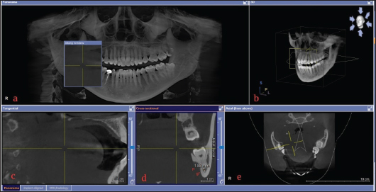
Figure 1. CBCT image for the investigation of the proximity of nerve (a) Panoramic view, (b) 3-D view, (c) Tangential view, (d) Cross-sectional image, f*to g*, the distance in millimeters, and (e) Axial plane.
Table 1. Relationship of age, gender, impaction status, and root status with the IANC proximity.
| Demographics | Frequency | Percentage | Proximity (mm ± SD) | p-value | |
|---|---|---|---|---|---|
| Age | 18-39 years | 6 | 4.5 | 1.45 ± 1.67 | 0.466* |
| 40-59 years | 112 | 83.6 | 1.80 ± 2.30 | ||
| 60 years and above | 16 | 11.9 | 2.51 ± 2.74 | ||
| Gender | Male | 76 | 56.7 | 2.44 ± 2.58 | 0.001 |
| Female | 58 | 43.3 | 1.13 ± 1.70 | ||
| Impaction type | Fully impacted | 112 | 83.6 | 1.46 ± 2.07 | 0.001 |
| Partially impacted | 22 | 16.4 | 4.53 ± 2.12 | ||
| Root status | Not fused | 73 | 54.5 | 2.21 ± 2.32 | 0.052 |
| Fused | 61 | 45.5 | 1.42 ± 2.37 | ||
p-values were obtained through the t-test and ANOVA*.
Table 2. Position of IANC and its proximity to the impacted third molar.
| Position of IANC | Frequency (n) | Percentage (%) | Proximity (mm ± SD) | p-value |
|---|---|---|---|---|
| Buccal | 36 | 26.9 | 0.77 ± 1.51 | <0.001 |
| Lingual | 14 | 10.4 | 0.31 ± 0.62 | |
| Apical | 75 | 56.0 | 2.91 ± 2.45 | |
| Inter-radicular | 5 | 3.7 | 0.00 ± 0.00 | |
| Superior | 4 | 3.0 | 0.00 ± 0.00 |
p-values were obtained through the chi-square.
Table 3. Gender-wise comparison of third molar impaction status, root status, and contact status of IANC.
| Variables | Total n (%) | Male n (%) | Female n (%) | p-value | |
|---|---|---|---|---|---|
| Winters classification | Disto-angular | 2 (1.5) | 0(0) | 2 (3.4) | 0.09 |
| Mesio-angular | 22 (16.4) | 9 (11.8) | 13 (22.4) | ||
| Horizontal | 21 (15.7) | 11 (14.5) | 10 (17.2) | ||
| Vertical | 89 (66.4) | 56 (73.7) | 33 (56.9) | ||
| Root status | Not fused | 73 (54.5) | 53 (69.7) | 19 (32.8) | <0.001 |
| Fused | 61 (45.5) | 22 (28.9) | 39 (67.2) | ||
| Contact status of IANC | No contact | 114 (85.1) | 45 (59.2) | 19 (32.8) | 0.02 |
| Contact | 20 (14.9) | 31 (40.8) | 39 (67.2) | ||
p-values were determined using the chi-square test.
According to Winter’s classification system, most of the impacted third molars were in a vertical position [89 (66.4%)] followed by mesioangular [22 (16.4%)] and horizontal [21 (15.7%)] positions. A significant correlation between gender and root fusion status, and contact of the IANC with the roots of the third molar is shown in Table 3.
Figure 2 illustrates fully impacted third molars in 62 (81.5%) males and 50 (86.2%) females and there was no discernible difference between both genders (p-value = 0.35).
Discussion
A number of factors contribute to the variation in IANC position among populations across the world, including genetic, developmental, and environmental differences. During the surgical removal of the lower wisdom teeth, the position of IANC is of critical importance. Furthermore, a lingually positioned or interradicular canal close to the lower wisdom tooth is more likely to cause an injury to the IAN.11
Gu et al.7 and Kim et al.12 reported that the majority of the Chinese (88.1%) and Korean (60%) population, respectively, had apically placed IANC, followed by buccally and lingual placement. Moreover, only 1% of the Korean population had IANC passing between the roots of the lower third molar.7,12 In a study conducted in Saudi Arabia, Shokry et al.13 reported that most of the individuals had inferiorly positioned IANCs, followed by lingual and buccal positions. In Odisha and the Netherlands, CBCT analysis revealed a majority of patients had IANC placed inferior to the impacted lower third molar.14,15 The findings of the present study are comparable to the studies conducted in the Korean and Saudi populations.
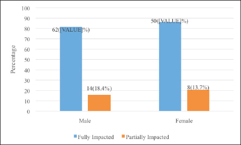
Figure 2. Distribution of third molar impaction type with respect to gender.
Various populations have different relationships between the IANC and the lower third molar. In a study on the Western Australian population, it was found that 88% of the subjects had a close proximity between the lower 3rd molar and the IANC (<1 mm) which is associated with a high risk of nerve injury during extraction.16 Previous studies conducted on the Korean, Indian, Nepali, and German populations found a close approximation of IANC to the roots of the lower 3rd molar in 28.3%, 67%, 46.8%, and 58% of the cases, respectively.17-20
Studies have supported the sexual dimorphism of the IANC canal, especially in the Southern and Eastern Indian populations.21,22 In addition, a study conducted in the United States reported that third molars were significantly closer to IANC in women than in men.23 This can be attributed to the different genetic, development, and dietary factors between males and females. However, studies conducted on Saudi Arabian, Nigerian, and Baltic populations have reported no significant difference between the males and females.24-26 There were no gender-specific differences in the distance between the IANC and the third molar in the present study.
Inconstant information on gender-wise comparison of lower 3rd molar impaction status in different countries is available. However, studies conducted in India and Iran by Hashemipour et al.27 and Padhye et al.28 reported a higher prevalence of third molar impaction in females. Regarding the impaction status of the lower wisdom, there was no difference between the genders in the current study.
Most of the participants in the current study had vertical impaction (Winter’s classification). Mesioangular impaction is the second most frequent impaction described in the literature, which is in coherence with the results of the present study.21 The vertical and mesio-angular patterns of angulation have statistically significant differences between females and males. The horizontal and disto-angular patterns in males remained statistically significant when compared to the same patterns in females.29 A study on the relationship between lower third molars and mandibular alveolar canal showed no association between winter classification and proximity of IANC.30
A proper understanding of the impact trend and position of IANC is very important to reach a better treatment and management plan in clinical settings. The implication of this knowledge will not only help in surgical planning and extraction but also in risk assessment, training, safe and effective dental procedures, forensic investigations, and diagnosing medical conditions.2,6
Conclusion
The close proximity between the IANC and the third molar roots emphasizes the potential risk for nerve injury during surgical extraction. For a variety of dental, medical, and forensic applications, it is crucial to understand the location of the IANC. There was a close relationship between the IAN and the third molar with the apically positioned canal having the least proximity to the third molar, especially in females and those with fully impacted third molars. Our findings highlight the importance of thorough radiographic analysis before any surgical procedure involving the impacted third molars.
Limitations of the study
The small sample size of the current study is one of the limitations. By employing random sampling and involving tertiary care hospitals, being the center for all types of populations, indiscriminate participation from the most representative subpopulation was ensured.
Acknowledgement
The authors would like to acknowledge the administration and focal persons of the participating institutes; Dr. Ali Shahid (Institute of Dentistry, CMH Lahore Dental College), Dr. Ijaz ur-Rehman (University College of Dentistry, Lahore), Dr. Muhammad Zahid Majeed (Shahida Islam Dental College, Lodhran), Dr. Muhammad Zeeshan (Faryal Dental College, Lahore).
List of Abbreviations
| CBCT | Cone beam computed tomography |
| IAN | Inferior alveolar nerve |
| IANC | Inferior alveolar nerve canal |
Conflict of interest
None to declare.
Grant support and financial disclosure
None to disclose.
Ethical approval
Ethical approval was granted by the IRB of the Institute of Dentistry, CMH Lahore Medical College, Lahore, Pakistan, vide Letter No. 599/ ERC/CMH/LMC, dated 25-05-2022.
Authors’ contributions
MA: Conception and design of the study, drafting of the manuscript, analysis and interpretation of data.
AN: Conception and design of the study, data collection, and drafting of the manuscript.
HH, KH, MT, SM: Data collection, analysis and interpretation of data, and drafting of the manuscript with critical intellectual input.
ALL AUTHORS: Approval of the final version of the manuscript to be published.
Authors’ Details
Mehtab Ahmad1, Abdullah Nasir2, Hammad Hassan3, Khalfan Haider1, Maliha Tariq1, Sonia Mubeen4
- Post Graduate Trainee, Periodontology Department, Institute of Dentistry, Combined Military Hospital Lahore Medical College, Institute of Dentistry, National University of Medical Sciences, Lahore, Pakistan
- Demonstrator, Community and Preventive Dentistry Department, Institute of Dentistry, Combined Military Hospital Lahore Medical College, Institute of Dentistry, National University of Medical Sciences, Lahore, Pakistan
- Assistant Professor, Dental Materials Department, University College Medicine of Dentistry, University of Lahore, Lahore, Pakistan
- Senior Registrar, Oral and Maxillofacial Surgery Department, Shahida Islam Medical and Dental College, Lodhran, Pakistan
References
- Deliverska EG, Petkova M. Complications after extraction of impacted third molars-literature review. J IMAB. 2016;22(3):1202–11. https://doi.org/10.5272/jimab.2016223.1202
- Shora KR, Channar KA, Shaikh IA, Memon AB, Shaikh AH, Maheshwari B, et al. Clinical presentation of mandibular impacted teeth and associated pathologies in the Unaizah, Al Qaseem; Saudi Arabia. J Pharm Res Int. 2021;33(19A):49–55. https://doi.org/10.9734/jpri/2021/v33i19A31327
- Qi W, Lei J, Liu YN, Li JN, Pan J, Yu GY. Evaluating the risk of post-extraction inferior alveolar nerve injury through the relative position of the lower third molar root and inferior alveolar canal. Int J Oral Maxillofac Surg. 2019 Dec;48(12):1577–83. https://doi: 10.1016/j.ijom.2019.07.008
- Bhangwar AW, Khan MI, Fatima H, Shams S. Inferior alveolar nerve injury assessment after surgical removal of mandibular third molar. Professional Med J. 2020;27(3):530–4. https://doi.org/10.29309/TPMJ/2020.27.3.3425
- Steel BJ, Surendran KSB, Braithwaite C, Mehta D, Keith DJW. Current thinking in lower third molar surgery. Br J Oral Maxillofac Surg. 2022;60(3):257–65. https://doi: 10.1016/j.bjoms.2021.06.016
- Zhao S, Wang Y, Yang X, Zhou X, Wang Z, Zhang K, et al. Extraction of impacted mandibular third molars in close proximity to the inferior alveolar canal with coronectomy-miniscrew traction to avoid nerve injury. Clin Oral Investig. 2023;27(8):4279–88. https://doi: 10.1007/s00784-023-05044-9
- Gu L, Zhu C, Chen K, Liu X, Tang Z. Anatomic study of the position of the mandibular canal and corresponding mandibular third molar on cone-beam computed tomography images. Surg Radiol Anat. 2018;40(6):609–14. https://doi.org/10.1007/s00276-017-1928-6
- Deppe H, Ritschl LM, Kleinschmidt J, Wagenpfeil S, Sculean A. Contiguity between the mandibular canal and the mandibular third molar in panoramic tomography compared with cone beam computed tomography: a topographic analysis. Quintessence Int. 2019;50(6):470–7. http://doi.org/10.3290/j.qi.a42485
- Santosh P. Impacted mandibular third molars: review of literature and a proposal of a combined clinical and radiological classification. Ann Med Health Sci Res. 2015;5(4):229–34. https://doi.org/10.4103/2141-9248.160177
- Maglione M, Costantinides F, Bazzocchi G. Classification of impacted mandibular third molars on cone-beam C.T. images. J Clin Exp Dent. 2015;7(2):e224–31. https://doi.org/10.4317/jced.51984
- Bhardwaj P, Bhardwaj Y, Ram R, Parmar M, Ghezta N, Sinha A. Radiographic factors associated with inferior alveolar nerve exposure during mandibular third molar surgery and their influence on neurosensory deficit: a prospective study. J Oral Biol Craniofac Res. 2022;12(6):818–22. https://doi: 10.1016/j.jobcr.2022.08.025
- Kim HJ, Jo YJ, Choi JS, Kim HJ, Kim J, Moon SY. Anatomical risk factors of inferior alveolar nerve injury association with surgical extraction of mandibular third molar in Korean population. Appl Sci (Basel). 2021;11(2):816. https://doi.org/10.3390/app11020816
- Shokry SM, Alshaib SA, Al Mohaimeed ZZ, Ghanimah F, Altyebe MM, Alenezi MA, et al. Assessment of the inferior alveolar nerve canal course among Saudis by cone beam computed tomography (pilot study). J Maxillofac Oral Surg. 2019;18(3):452–8. https://doi.org/10.1007/s12663-018-1167-3
- Mohanty R, Rout P, Singh V. Preoperative anatomic evaluation of the relationship between inferior alveolar nerve canal and impacted mandibular third molar in a population of Bhubaneswar, Odisha, using CBCT: a hospital-based study. J Maxillofac Oral Surg. 2020;19(2):257–62. https://doi.org/10.1007/s12663-019-01193-1
- Ghaeminia H, Meijer GJ, Soehardi A, Borstlap WA, Mulder J, Bergé SJ. Position of the impacted third molar in relation to the mandibular canal. Diagnostic accuracy of cone beam computed tomography compared with panoramic radiography. Int J Oral Maxillofac Surg. 2009;38(9):964–71. https://doi.org/10.1016/j.ijom.2009.06.007
- Winstanley KL, Otway LM, Thompson L, Brook ZH, King N, Koong B, et al. Inferior alveolar nerve injury: correlation between indicators of risk on panoramic radiographs and the incidence of tooth and mandibular canal contact on cone‐beam computed tomography scans in a Western Australian population. J Investig Clin Dent. 2018;9(3):e12323. https://doi.org/10.1111/jicd.12323
- Lee B, Park Y, Ahn J, Chun J, Park S, Kim M, et al. Assessment of the proximity between the mandibular third molar and inferior alveolar canal using preoperative 3D-CT to prevent inferior alveolar nerve damage. Maxillofac Plast Reconstr Surg. 2015;37(1):30. https://doi.org/10.1186/s40902-015-0030-4
- KalaiSelvan S, Ganesh SKN, Natesh P, Moorthy MS, Niazi TM, Babu SS. Prevalence and pattern of impacted mandibular third molar: an institution-based retrospective study. J Pharm Bioallied Sci. 2020;12(Suppl 1):S462–7. https://doi.org/10.4103/jpbs.JPBS_140_20
- Nyachhyon R, Joshi U, Mainali A, Sakya P. Compression of the inferior alveolar canal by mandibular third molar among images taken from patients visiting dental imaging centers of Kathmandu: a descriptive cross-sectional study. JNMA J Nepal Med Assoc. 2022;60(245):26–30. https://doi.org/10.31729/jnma.7124
- Kuntz NM, Schulze R. Three-dimensional classification of lower third molars and their relationship to the mandibular canal. J Oral Maxillofac Surg. 2021;79(8):1611–20. https://doi.org/10.1016/j.joms.2021.02.033
- Sailaja S, Shil M, Lavanya R, Ravali T, Fathima P, Aparna T. Significance of mandibular canal position and its foramina in cone beam computed tomography images of the mandible for analyzing sexual dimorphism- a retrospective study. J Indian Acad Oral Med Radiol. 2022;34(3):338. https://doi.org/10.4103/jiaomr.jiaomr_288_21
- Rath R, Sangamesh NC, Acharya RR, Sharma G. Sexual dimorphism of inferior alveolar canal location: a record- based CBCT study in Eastern India. J Oral Maxillofac Pathol. 2022 Apr-Jun;26(2):277–82. https://doi: 10.4103/jomfp.jomfp_139_21
- Simonton JD, Azevedo B, Schindler WG, Hargreaves KM. Age- and gender-related differences in the position of the inferior alveolar nerve by using cone beam computed tomography. J Endod. 2009 Jul;35(7):944–9. https://doi:10.1016/j.joen.2009.04.032
- Zaman MU, Almutairi NS, Abdulrahman Alnashwan M, Albogami SM, Alkhammash NM, Alam MK. Pattern of mandibular third molar impaction in nonsyndromic 17760 patients: a retrospective study among Saudi population in central region, Saudi Arabia. Biomed Res Int. 2021;2021:1880750. https://doi.org/10.1155/2021/1880750
- Osunde O, Bassey G. Pattern of impacted mandibular third molars in Calabar, Nigeria. Afr J Med Health Sci. 2016;15(1):14. https://doi.org/10.4103/2384-5589.183886
- Jaroń A, Trebek G. The pattern of mandibular third molar impaction and assessment of surgery difficulty: a retrospective study of radiographs in east Baltic population. Int J Environ Res Public Health. 2021;18(11):6016. https://doi.org/10.3390/ijerph18116016
- Hashemipour MA, Tahmasbi-Arashlow M, Fahimi-Hanzaei F. Incidence of impacted mandibular and maxillary third molars: a radiographic study in a Southeast Iran population. Med Oral Patol Oral Cir Bucal. 2013;18(1):e140–5. https://doi.org/10.4317/medoral.18028
- Padhye MN, Dabir AV, Girotra CS, Pandhi VH. The pattern of mandibular third molar impaction in the Indian population: a retrospective clinical-radiographic survey. Oral Surg Oral Med Oral Pathol Oral Radiol. 2013;116(3):e161–6. https://doi.org/10.1016/j.oooo.2011.12.019
- Kumar SM, Al-Hobeira H, Shaikh S, Siddiqui AA, Syed J, Mian RI. Distribution of impacted third molars based on gender and patterns of angulation in dental students of the Hai’l region, Saudi Arabia: a panoramic radiographic (OPG) study. Int J Contemp Med Res. 2017;4(9):1829–32.
- Armijos Salinas CA, González Bustamante AM, Quel Carlosama FE. Relationship between lower third molars and mandibular alveolar canal through cone beam CT scans. Univ Odontol. 2021;40:1–14. https://doi.org/10.11144/Javeriana.uo40.rltm