Case Report
VOLUME: 37 | ISSUE: 4 | Dec 25, 2021 | PAGE: (201 - 206) | DOI: 10.51441/BioMedica/5-608
Pediatric craniopharyngioma with a rare presentation of tooth enamel like structures - a case report
Authors: Fizza Waqar , Mahvish Hussain , Shazia Riaz , Amber Goraya , Laeeq Ahmad , Samina Zaman
Article Info
Authors
Fizza Waqar
Department of Histopathology, Children Hospital and the University of Child Health Sciences, Lahore, Pakistan
Mahvish Hussain
Department of Histopathology, Children Hospital and the University of Child Health Sciences, Lahore, Pakistan
Shazia Riaz
Department of Haemoncology, Children Hospital and the University of Child Health Sciences, Lahore, Pakistan
Amber Goraya
Department of Radiology, Children Hospital and the University of Child Health Sciences, Lahore, Pakistan
Laeeq Ahmad
Department of Neurosurgery, Children Hospital and the University of Child Health Sciences, Lahore, Pakistan
Samina Zaman
Department of Histopathology, Children Hospital and the University of Child Health Sciences, Lahore, Pakistan
Publication History
Received: October 11, 2021
Revised: November 19, 2021
Accepted: December 18, 2021
Published: December 25, 2021
Abstract
Craniopharyngioma (CP) is a rare tumor accounting for <1% of all primary central nervous system (CNS) tumors. We herein report a case of a 2.5 years old male child diagnosed with supra-sellar tumor on prenatal ultrasound. Histologically, the tumor had tooth enamel like structures, one of the rarest finding in the literature. This case from Pakistan is hereby reported as a seventh such case world-wide. Our patient is a rare presentation of an antenatal diagnosis of CP with World Health Organization (WHO) grade I.
Keywords: Craniopharyngioma, tumor, pediatric, central nervous system (CNS), tooth enamel.

Pubmed Style
Fizza Waqar, Mahvish Hussain, Shazia Riaz, Amber Goraya, Laeeq Ahmad, Samina Zaman. Pediatric craniopharyngioma with a rare presentation of tooth enamel like structures - a case report. BioMedica. 2021; 25 (December 2021): 201-206. doi:10.51441/BioMedica/5-608
Web Style
Fizza Waqar, Mahvish Hussain, Shazia Riaz, Amber Goraya, Laeeq Ahmad, Samina Zaman. Pediatric craniopharyngioma with a rare presentation of tooth enamel like structures - a case report. https://biomedicapk.com/articles/online_first/608 [Access: July 03, 2024]. doi:10.51441/BioMedica/5-608
AMA (American Medical Association) Style
Fizza Waqar, Mahvish Hussain, Shazia Riaz, Amber Goraya, Laeeq Ahmad, Samina Zaman. Pediatric craniopharyngioma with a rare presentation of tooth enamel like structures - a case report. BioMedica. 2021; 25 (December 2021): 201-206. doi:10.51441/BioMedica/5-608
Vancouver/ICMJE Style
Fizza Waqar, Mahvish Hussain, Shazia Riaz, Amber Goraya, Laeeq Ahmad, Samina Zaman. Pediatric craniopharyngioma with a rare presentation of tooth enamel like structures - a case report. BioMedica. (2021), [cited July 03, 2024]; 25 (December 2021): 201-206. doi:10.51441/BioMedica/5-608
Harvard Style
Fizza Waqar, Mahvish Hussain, Shazia Riaz, Amber Goraya, Laeeq Ahmad, Samina Zaman (2021) Pediatric craniopharyngioma with a rare presentation of tooth enamel like structures - a case report. BioMedica, 25 (December 2021): 201-206. doi:10.51441/BioMedica/5-608
Chicago Style
Fizza Waqar, Mahvish Hussain, Shazia Riaz, Amber Goraya, Laeeq Ahmad, Samina Zaman. "Pediatric craniopharyngioma with a rare presentation of tooth enamel like structures - a case report." 25 (2021), 201-206. doi:10.51441/BioMedica/5-608
MLA (The Modern Language Association) Style
Fizza Waqar, Mahvish Hussain, Shazia Riaz, Amber Goraya, Laeeq Ahmad, Samina Zaman. "Pediatric craniopharyngioma with a rare presentation of tooth enamel like structures - a case report." 25.December 2021 (2021), 201-206. Print. doi:10.51441/BioMedica/5-608
APA (American Psychological Association) Style
Fizza Waqar, Mahvish Hussain, Shazia Riaz, Amber Goraya, Laeeq Ahmad, Samina Zaman (2021) Pediatric craniopharyngioma with a rare presentation of tooth enamel like structures - a case report. , 25 (December 2021), 201-206. doi:10.51441/BioMedica/5-608
Biomedica - Official Journal of University of Health Sciences, Lahore, Pakistan
Volume 37(4):201-206
CASE SERIES
Pediatric craniopharyngioma with a rare presentation of tooth enamel like structures - a case report
Fizza Waqar1*, Mahvish Hussain1, Shazia Riaz2, Amber Goraya3, Laeeq Ahmad4 and Samina Zaman1
Received: 11 October 2021 Revised date: 19 November 2021 Accepted: 18 December 2021
Correspondence to: Fizza Waqar
*Department of Histopathology, Children Hospital and the University of Child Health Sciences, Lahore, Pakistan.
Email: doctorfkhan@gmail.com
Full list of author information is available at the end of the article.
ABSTRACT
Craniopharyngioma (CP) is a rare tumor accounting for <1% of all primary central nervous system tumors. We herein report a case of a 2.5 years old male child diagnosed with supra-sellar tumor on prenatal ultrasound. Histologically, the tumor had tooth enamel like structures, one of the rarest finding, in the literature. This case from Pakistan is hereby reported as a seventh such case world-wide. Our patient is a rare presentation of an antenatal diagnosis of CP with World Health Organization grade I.
Keywords:
Craniopharyngioma, tumor, pediatric, central nervous system (CNS), tooth enamel.
Introduction
Craniopharyngioma (CP) is accounted as one of the rare tumours (<1%) of the central nervous system with predominance in pediatric age group1. Jakob Erdheim, a pathologist from Austria, for the first time in 1904 reported its origin from squamous cell rests present in the remnant ducts of the hypophyses and pharynx encasing the infundibulum and supra-sellar regions with extension up to the hypothalamus, third ventricle, optic chiasma, and infiltration of cranial nerves.2,3 The most frequently reported sites of CP in the brain remains the sellar and suprasellar regions.4,5
Case Presentation
Our patient, now approximately two and a half years old boy, was initially diagnosed on routine prenatal ultra sound at 23.3 weeks of gestation with 1.6 × 1.4 cm echogenic mass in the interpedencular cistern (Figure 1). The baby was delivered at 40 weeks of gestation by a planned caesarean section. At birth, the baby had no abnormality on body surface. He was moving all four limbs equally and feeding well. He achieved normal mile stones till the age of 6 months. However, around 9 months of age, parents noticed a rapid enlargement of his head, and he was unable to stand without support.
The computerized tomography scan at that time revealed enlargement in size of the mass which measured 5.2 × 4.6 × 4.7 cm and located at the supra-sellar region (Figure 2). Clinical diagnosis of CP was made. The surgery was postponed till the patient was 2 years of age. Magnetic resonance imaging (MRI) at the time of surgery showed approximately 6.7 × 6.2 × 5.7 cm supra-sellar and interpeduncular heterogenous mass with coarse calcifications (Figures 3 and 4).
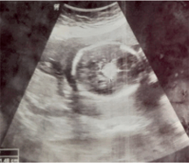
Figure 1. Ultrasound showing approximately 1.6 × 1.4 cm sized echogenic mass in interpeduncular cistern raising suspicion of brain tumour at gestational age of 23 weeks and 3 days.
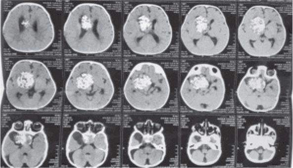
Figure 2. CT Brain Plain showing supra-sellar and interpenduncular heterogenous mass with coarse calcifications measuring approximately 5.2 × 4.6 × 4.7 cm.
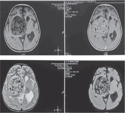
Figure 3. a: TW1 MRI Pre & Post Contrast supra-sellar mass measuring 6.7 × 6.2 × 5.7 cm. b: TW2 & diffusion-weighted imaging Axial MRI.
At the age of 2 years, right parietal craniotomy was performed in October 2020. The excised mass revealed a rare spectacle of pronounced odontogenic elements / tooth formation on both gross appearance as well as on histological examination. The routine CPs on gross analysis have cysts with contents either like dark green-brown machine oil or having clear fluid. In contrast the specimen in our case showed multiple grey red to greyish pieces of tissue with many pearly white tooth-like structures (Figure 5). Histologically, the sections revealed nodules of plump anucleated squamous cells, ghost cells, wet keratin along with large foci of dystrophic calcification. Moreover, odontogenic rests and obvious odontogenic differentiation was present revealing multiple malformed teeth. On these histological findings, the diagnosis of CP World Health Organization (WHO) grade I with tooth like structures was rendered (Figures 6-8). Postoperative MRI showed a minimal residual mass in sellar/ suprasellar region. However, the patient is clinically well and had no post-operative complications till 6 months after the surgery.
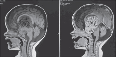
Figure 4. TW1 pre & post contrast sagittal MRI.
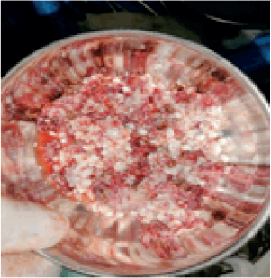
Figure 5. Multiple gray white and red tumor pieces with intervening pearly white tumor nodules resembling teeth structures.
Discussion
CP is a benign but locally invasive tumor of the sellar region.6 Despite the benign histological grade I classification, CP often recurs and may cause severe morbidity due to its close anatomic relation with important visual and endocrinological structures.1,4,7,8 Impaired visual function is a primary manifestation in 23%-58% of all children diagnosed with a CP.4
On MRI, these tumors often show T1WI bright cyst due to their high protein content and the solid components usually demonstrate heterogenous signals on T1-weighted (TW1) as well as T2-weighted (TW2).9 Histologically, the commonly reported pediatric variant is adamantinomatous (aCP) tumour while papillary subtype (pCP) is typically seen in adults.6,9 Possible origin of this type of CP is from a partial obliteration or metaplastic changes in the craniopharyngeal duct while some authors hypothesize inrolling of the dentine and similar structures to the developing pituitary gland. Its unique similarity to the adamantinoma of the jaw may mark its potential link with the embryonic ductal epithelium.9
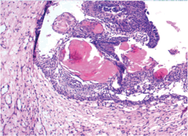
Figure 6. Photomicrograph showing CP adamantinomatous type comprising of ghost cells with peripheral palisading (H & E × 200).
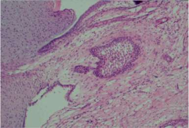
Figure 7. Photomicrograph showing loose stellate reticulum (H & E × 200).
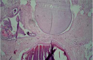
Figure 8. Photomicrograph showing primitive odontogenic mesenchyme and epithelium with rudimentary tooth formation, dentine and enamel (H & E × 100).
The reported incidence of CP is 0.5-2.0 new patients per million persons per year.7,8 The two peaks in the age distribution of the tumor reported in literature show <10 and >50 years of age.1,5,7,9,10
The most commonly presenting symptoms are vision disturbances, high intracranial pressure and endocrine manifestations related to the dysfunction of hypothalamus and/or pituitary gland.1,2,4,5,7,8,9 It is rarely reported during the fetal life with a scant published data in the neonatal period. We present the case of a patient who got diagnosed with the supra-sellar tumor on prenatal ultrasound and had surgical resection after birth showing tooth-like enamel structures on a histological diagnosis. Only six cases of CP with tooth formation have been reported till now worldwide.9
Conclusion
The presentation of the current study is a rare presentation of antenatal diagnosis of CP WHO grade I with tooth like structures having successful surgical resection. The differential diagnosis of CP with tooth like structures includes teratoma. The intimate embryological origin of tumors rising from Rathkes Pouch should be kept in mind when detecting tumors in the sellar / suprasellar region.
Limitations of the case report
As of any case report, the causal relationship could not be established. Being in neonatal period, endocrine and visual disturbances of the patient could not be ascertained in a progressive manner till follow-up.
Acknowledgement
The authors acknowledge all the staff members of the Departments of Histopathology, Haemoncology, Radiology and Neurosurgery, Children Hospital and the University of Child Health Sciences, Lahore, Pakistan who helped in the acquisition of data for this manuscript.
List of Abbreviations
| CP | Craniopharyngioma |
| MRI | Magnetic resonance imaging |
| TW1 | T1-weighted |
| TW2 | T2-weighted |
| WHO | World Health Organization |
Conflict of interest
None to declare.
Grant support and financial disclosure
None to disclose.
Ethical approval
Ethical approval has been taken by the Ethics Committee of The Children Hospital and the University of Child Health Sciences, Lahore, Pakistan vide reference letter number IRB-547/2021 dated 10/02/2021.
Authors’ contribution
FW, MH & SZ: Conception of study, drafting of manuscript, critical revision with important intellectual content.
SR & AG: Drafting of manuscript.
LA: Acquisition of data.
ALL AUTHORS: Approval of the final version of the manuscript to be published.
Authors’ details
Fizza Waqar1, Mahvish Hussain1, Shazia Riaz2, Amber Goraya3, Laeeq Ahmad4 and Samina Zaman1
- Department of Histopathology, Children Hospital and the University of Child Health Sciences, Lahore, Pakistan
- Department of Haemoncology, Children Hospital and the University of Child Health Sciences, Lahore, Pakistan
- Department of Radiology, Children Hospital and the University of Child Health Sciences, Lahore, Pakistan
- Department of Neurosurgery, Children Hospital and the University of Child Health Sciences, Lahore, Pakistan
References
- Jensterle M, Jazbinsek S, Bosnjak R, Popovic M, Zalete LZ, Vesnaver TV, et al. Advances in the management of craniopharyngioma in children and adults. Radiol Oncol. 2019;53(4):388–96. https://dx.doi.org/10.2478%2Fraon-2019-0036
- Pascual JM, Prieto R, Rosdolsky M, Strauss S, Castro I, Verena D. Cystic tumors of the pituitary infundibulum: seminal autopsy specimens (1899 to 1904) that allowed clinical-pathological craniopharyngioma characterization. Pituitary. 2018;21(4):393–405. https://doi.org/10.1007/s11102-018-0889-z
- Zada G, Lin N, Ojerholm E, Ramkissoon S, Laws ER. Craniopharyngioma and other cystic epithelial lesions of the sellar region: areviewofclinical,imaging, andhistopathological relationships. Neurosurg Focus. 2010;28(4):E4. https://doi.org/10.3171/2010.2.FOCUS09318
- Huang CC, Lin KL, Wu CT, Jung SM, Wang CJ, Chen YC, et al. Clinical and endocrinological manifestations of childhood-onset craniopharyngioma before surgical removal: a report from one medical center in Taiwan. Pediatr Neonatol. 2021;62(2):181–6. https://doi.org/10.1016/j.pedneo.2020.08.014
- Ramanbhavana VS, Vara Prasad KS. A case series of craniopharyngioma: epidemiological study and management analysis at tertiary care center. Asian J Neurosurg. 2019;14(4):1196–202. https://doi.org/10.4103/ajns.ajns_67_19.
- Larkin JS, Olaf A. Pathology and pathogenesis of craniopharyngiomas. Pituitary. 2013;16(1):9–17. https://doi.org/10.1007/s11102-012-0418-4
- Müller HL. Craniopharyngioma. Handb Clin Neurol. 2014;124:235–53. https://doi.org/10.1016/b978-0-444-59602-4.00016-2
- Müller H. Childhood craniopharyngioma - current concepts in diagnosis, therapy and follow-up. Nat Rev Endocrinol. 2010;(6):609–18. https://doi.org/10.1038/nrendo.2010.168
- Muller C, Adroos, N, Lockhat, Z, Tomas Slavik T, Kruger H. Toothy craniopharyngioma: a literature review and case report of craniopharyngioma with extensive odontogenic differentiation and tooth formation. Childs Nerv Syst. 2011;27(2):323–6. https://doi.org/10.1007/s00381-010-1296-6
- Manet R, Apra C, Jouanneau E. Epidemiology, clinical presentation, and prognosis of adult-onset craniopharyngioma. In: Adult Craniopharyngiomas. Springer; 2020. https://doi.org/10.1007/978-3-030-41176-3_3