Case Report
VOLUME: 37 | ISSUE: 3 | Sep 25, 2021 | PAGE: (148 - 150) | DOI: 10.51441/BioMedica/5-505
A Case of Unilateral Neonatal Mastauxe
Authors: Muhammad Imran , Mina Amer , Aqsa Majeed , Uqba Afzal
Article Info
Authors
Muhammad Imran
Department of Pathology, Allama Iqbal Medical College, Lahore-Pakistan.
Mina Amer
Department of Pathology, Allama Iqbal Medical College, Lahore-Pakistan.
Aqsa Majeed
Department of Pathology, Allama Iqbal Medical College, Lahore-Pakistan.
Uqba Afzal
Department of Pathology, Allama Iqbal Medical College, Lahore-Pakistan.
Publication History
Received: April 11, 2021
Revised: July 19, 2021
Accepted: August 08, 2021
Published: September 25, 2021
Abstract
Enlargement of breast tissue in neonates is a condition seen as a physiological response to the decline in maternal hormonal levels postnatal. It is commonly seen in the initial weeks, but can progress in size within two months of life. We herein report a case diagnosed as neonatal mastauxe in a two months old female, which was followed up without any medical or surgical intervention. Neonatal mastauxe means enlargement of breast tissue in newborn, which is sometimes essential to be differentiated from breast abscess and mastitis in babies.
Keywords: Neonate, Breast, Enlargement, Physiological, Mastauxe

Biomedica - Official Journal of University of Health Sciences, Lahore, Pakistan
Volume 37(3):148-150
CASE REPORT
A case of unilateral neonatal mastauxe
Muhammad Imran1*, Mina Amer1, Aqsa Majeed1, Uqba Afzal1
Received: 11 April 2021 Revised date: 19 July 2021 Accepted: 08 August 2021
Correspondence to: Muhammad Imran
*Department of Pathology, Allama Iqbal Medical College, Lahore, Pakistan.
Email: drelmo@hotmail.com
Full list of author information is available at the end of the article.
ABSTRACT
Enlargement of breast tissue in neonates is a condition seen as a physiological response to the decline in maternal hormonal levels postnatal. It is commonly seen in the initial weeks, but can progress in size within 2 months of life. We herein report a case diagnosed as neonatal mastauxe in a 2-monthold female, which was followed up without any medical or surgical intervention. Neonatal mastauxe means enlargement of breast tissue in newborn, which is sometimes essential to be differentiated from breast abscess and mastitis in babies.
Keywords:
Neonate, breast, enlargement, physiological, mastauxe.
Introduction
Breast enlargement in neonates is a usual pediatric presentation that can be seen mostly in the initial weeks of life, but can show increase in the size in the next 2 months.1 Neonatal breast enlargement is reported in almost 65%-90% of the infants in their neonatal lives. Physiologically the declining levels of maternal hormones, especially estrogen in the last weeks of pregnancy, which gives feedback for the release of prolactin from the pituitary gland of the neonate.2 In some cases, galactorrhea can be an accompanying presentation and the enlarged breast tissue secretes whitish liquid. It is called “witch’s milk” and is known to show similar composition as mother’s milk.3 Neonatal mastauxe is a term used for the enlargement of breast in the neonates. It is presents as bilateral swellings, with increase in size over 2 months. It is a physiologic self-limiting condition which requires no medical or surgical treatment.2 At times, antibiotic management is essential if there is associated mastitis or formation of a breast abscess.3
We herein report a case of unilateral breast enlargement in a 2-month-old female child who was brought to the outpatient department of the hospital. She was investigated, diagnosed, and followed up without any major intervention.
Case Presentation
A 2-month-old female child presented in the outpatient department of the Jinnah Hospital, Lahore, Pakistan with enlargement of the right breast. She was born by normal vaginal delivery at 37 weeks of an uncomplicated gestation. She was examined by the neonatologist at the time of birth. There were no congenital abnormalities. Neonatal jaundice was not present. The parents of the infant noticed slight bilateral breast swellings in the first month after birth. They ignored the swelling, considering it a part of the neonatal physique as the baby was healthy. The right breast started increasing in size slowly in the second month of life (Figure 1). Initially, some home remedies were tried to reduce the swelling, including rubbing, squeezing, and oil massage with olive oil. According to the parents, there was no watery or purulent discharge since the start of the swelling. There was no history of associated pyrexia. Parents were worried about their child as they were misled by the people that breast lump means breast cancer.
The chest examination of the female revealed enlarged right breast, measuring 5.0 cm × 4.0 cm in size. The left breast was also slightly swollen, measuring 2.0 cm × 1.0 cm. The nipple on the right side was slightly depressed because of the swelling. On palpation, the right breast was soft to firm. There was no discharge on pressing the nipple in bilateral breasts. No signs of inflammation were noted. Laboratory tests were conducted. Complete blood count was within normal limits. Grey-scale ultrasonography showed an oval mass 43.0 mm × 22.0 mm in the right breast and a 9.0 mm × 8.0 mm in the left breast. There were small non-echogenic cystic spaces in the right breast with no pus collection. The radiological evaluation was reported as benign breast enlargement. Prolactin levels in the female revealed an increase up to 42.1 ng/ml (normal range, 2.5-12.8 ng/dl); however, the levels of follicle-stimulating hormone, luteinizing hormone, estrogen, and progesterone were with reference range.
Taking the apprehension of the parents into account, fine needle aspiration cytology (FNAC) of the right breast swelling was performed under aseptic conditions in the Pathology Department. The smears were air dried and ethanol fixed. The slides were stained with Hematoxylin & Eosin and Giemsa stains. The aspirates from FNAC revealed few scattered groups of ductal cells with regular nuclei. No atypia was identified. No evidence of infection was noted (Figure 2). The parents were counseled that the condition is physiological and no intervention is need. The female was discharged without any medical treatment. Follow up of the female was scheduled after every 2 to 3 months.
Discussion
Breast enlargement in the neonates is a common physiologic phenomenon produced by the decreased levels of maternal estrogen which in turn elevates the prolactin levels as feedback to the pituitary gland in neonates.1 The breast enlargement is usually unilateral and may present with increase in size for up to 2 months. Bilateral enlargement may also be seen. Hypersensitivity of the neonatal breast tissue to estrogen and prolactin is an important factor for this enlargement; however, the variability in response to hormones in neonates is still not clear with definite reasons.2
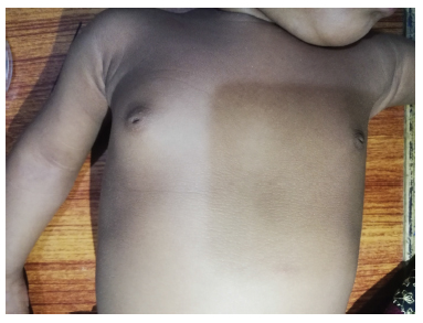
Figure 1. 2-month old female showing right sided breast tissue enlargement.
Mastauxe comes from Greek words “mastos” and “auxein”, meaning breast and enlargement, respectively. Neonatal mastauxe refers to a physiologic enlargement of breast in neonates, which is less than 3 cm in size. Giant neonatal mastauxe is defined when the size of the swelling is more than 3 cm.1 Inflammation of the breast tissue in neonates is termed as neonatal mastitis, which involves superadded infectious process, and formation of pus collection is referred to as neonatal breast abscess. Both these clinical conditions need antibiotic treatment.4
Neonatal mastauxe is seen in 65%-90% of newborns, which usually occurs in early weeks after birth and subsides within few weeks. A 2-week-old female baby, who presented with bilateral enlargement of breast with tenderness and whitish discharge from nipples, was treated with intravenous antibiotic therapy.4 Another baby girl presented with bilateral breast swellings in her first week of life. There is no laterality predilection and the condition is independent of the gender. In our case, a female infant presented with a unilateral swelling at the end of her 4 weeks life and as expected, the serum prolactin levels were raised.5
It is important to differentiate between mastitis and breast abscess. Staphylococcus aureus is the most common micro-organism known to cause abscess in the breast. Ultrasonography is a non-invasive radiologic technique to diagnose inflammation or any collection of pus in the enlarged breast tissue. In our case, inflammation or infection was not present. Fine needle aspiration was performed, considering unilaterality of the condition, and also to release the apprehension of the parents regarding any malignancy.6 The management of the neonatal mastauxe is follow-up with observation to look for regression. The enlargement regresses spontaneously within weeks. It is very important to properly educate the parents about this physiologic condition, as squeezing and pressing may exaggerate the swelling, with chances of inflammation and infection, leading to abscess formation. Also, long-term follow-up of the patients with this condition is recommended, especially in the female gender because of the hypersensitivity to estrogen, which may make breast susceptible for atypical changes in the adult life.7
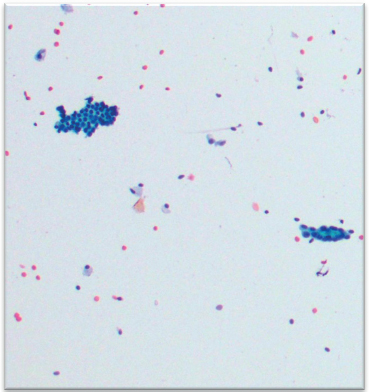
Figure 2. FNAC showing normal breast cells and no inflammation.
Conclusion
Neonatal mastauxe is a benign, physiologic condition which regresses spontaneously. Ultrasonography may clearly differentiate inflammation or an abscess. Follow-up of the neonate with observation is the only management apart from parental reassurance that the swelling will regress spontaneously within few weeks. Parents’ education is mandatory to avoid mishandling and chances of infection.
Limitations of the study
The neonate was followed for 2 months in the hospital, which showed gradual regression in the size of breast tissue. The family belongs to another city which makes it difficult for them to attend hospital visits every month. The parents were then followed up for another few weeks telephonically and on image observations it was revealed that the baby is healthy and there are no further complaints.
Acknowledgement
The authors acknowledge Dr. Naeem Khan in Radiology Department for providing the valuable comments on ultrasonography, Dr. Haseeb Mehmood from Surgery Department and Dr. Tazeen Anis and Dr. Nusrat Zahra from Pathology Department for their input in the case report.
List of Abbreviations
| FNAC | Fine needle aspiration cytology. |
Conflict of interest
None to declare.
Grant support and financial disclosure
None to disclose.
Ethical approval
The written informed consent was taken from the parents. The photograph of the neonate was taken and submitted by her father himself. Approval was taken from Institutional Review Board (Allama Iqbal Medical College, Lahore, Pakistan) on 11th February 2021, numbered AIMC/02/2021.
Author’s contribution
MI: Conception of study, drafting of manuscript, critical revision with important intellectual content; MA, AM: Drafting of manuscript; UA: Acquisition of data; ALL AUTHORS: Approval of the final version of the manuscript to be published.
Authors’ details
Muhammad Imran1*, Mina Amer1, Aqsa Majeed1, Uqba Afzal1
Department of Pathology, Allama Iqbal Medical College, Lahore, Pakistan
References
- Raveenthiran V. Neonatal mastauxe (breast enlargement of the newborn). J Neonatal Surg. 2013;2(3):31–4. https://doi.org/10.47338/jns.v2.43
- Suthar B, Aggarwal K. A rare case of neonatal mastauxe: case report. Indian J Radiol Imaging. 2019;29(1):89–93. https://doi.org/10.4103/ijri.IJRI_317_18
- Weinstein SP, Conant EF, Orel SG, Zuckerman JA, Bellah R. Spectrum of US findings in pediatric and adolescent patients with palpable breast masses. Radiographics. 2000;20(6):1613–21. https://doi.org/10.1148/radiographics.20.6.g00nv091613
- Donaire A, Guillen J, Rajegowda B. Neonatal breast hypertrophy: revisited. Pediatr Ther. 2016;6(4):297–301. https://doi.org/10.4172/2161-0665.1000297
- Cameselle-Teijeiro JF, Mallo R, Otero M. Alterations in the development of the breast newborn: Giant mastauxe (exaggerated form of neonatal mastauxe). Open J Clin Med Case Rep. 2016;2(1):1197.
- Meshram GG, Kaur N, Hura KS. Breast enlargement in newborns: a folkloric-medical dilemma. Trop Dr. 2018;48(2):150–1. https://doi.org/10.1177/0049475517729068
- Valerio E, Palatron S, Vanzo V, Vendramin S, Cutrone M. Breast disorders of the newborn. Arch Dis Child Educ Pract Ed. 2016;101(5):236–8. https://doi.org/10.1136/archdischild-2016-310881